INSIDE AN ANIMAL · ORIGINAL 3D STUDY
Why do a cat’s eyes shine—but not make their own light?
A second chance to catch the light.
Take it apart and inspect every system.
Click a part to highlight it and read why it matters.
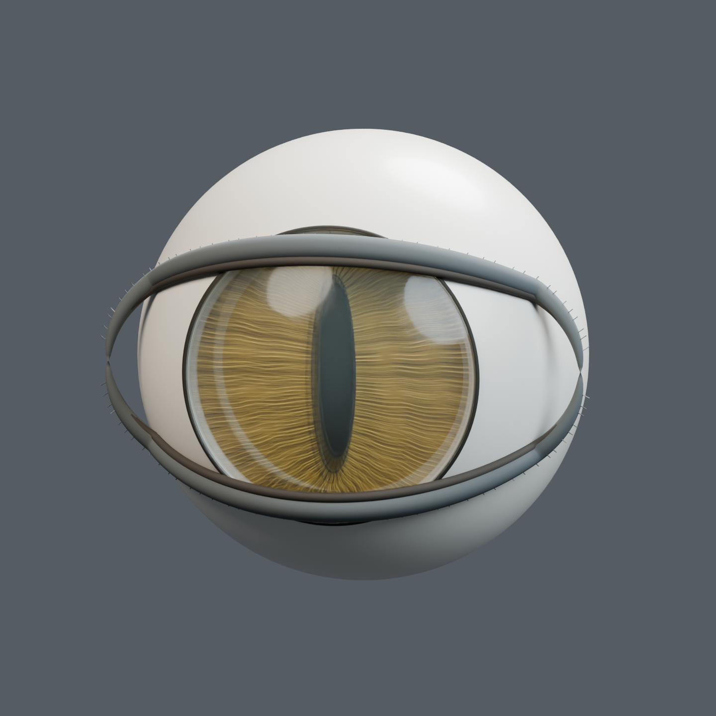
A familiar object. A hidden system.
A second chance to catch the light.
Eyeshine is borrowed light.
The eye collects light from outside. Some light that passes through the retina reaches a reflective region called the tapetum, then passes through the retina again. Separate the nested tissues to find that hidden reflector.
The tapetum returns incoming light. It does not make its own.
Read the explanation
The pupil is an opening—not a black plug.External light passes through the clear cornea and the opening in the iris. The lens sits behind the iris. This narrow vertical pupil is one illustrative state; this model does not animate dilation.
The retina detects light.The cornea and lens help focus light onto the retina. Retinal cells convert light into neural information. The optic nerve carries that information toward the brain—it is not a tube carrying light.
The reflector is inside the choroid.The tapetum is a localized reflective region within the choroid, behind the retina. It returns some previously unabsorbed light through the retina, giving it another opportunity to be detected. It is not a complete glowing lining. A teaching window exposes the otherwise covered layers.
No incoming light. No eyeshine.Some reflected light can leave the eye toward an observer, producing eyeshine. It is reflection, not light made by the eye. Switch off the incoming-light markers: both inward and return paths disappear. The anatomical model stays lit so you can inspect it; this is not a simulation of seeing in total darkness.
Sources and safety
- Cornell University College of Veterinary Medicine: The eye transmits and focuses incoming light onto the retina, where detection leads to neural information carried toward the brain.
- Cornell University College of Veterinary Medicine: The tapetum reflects some light back through the retina, providing a second opportunity for detection rather than creating new light.
- University of Minnesota: The tapetal region lies within the choroid, behind the retina, and is localized rather than covering the whole eye.
Do not shine lasers or intense lights into an animal’s eyes. This is not veterinary advice or a way to diagnose eye disease. Concerns about vision or eye appearance belong with a veterinarian.
Scope and limitations
A generic domestic-cat eye explaining external light, retinal detection and a localized tapetum. This is a macro-anatomy teaching model, not a scan, histological reconstruction, diagnostic model or optical simulation.
- Original generic feline eye, not a scan, medical device or veterinary diagnosis.
- Spatial decomposition shows nested tissue relationships, not literal disassembly.
- Anatomical layer thicknesses, colours and spacing are simplified for readability; dimensions are illustrative.
- Tapetum is a localized dorsal/posterior specialization within the choroid, behind the retina; no emissive anatomical materials are used.
- The retinal viewing window is an explicit cutaway device, not an anatomical hole.
- Pupil is an aperture in this narrow vertical state; the tapetal region does not expand with pupil dilation.
- Light paths, if shown, must be schematic and stop when the external light source is off. The optic nerve carries neural signals, not light.
- The model is lit for inspection even when the external-light markers are switched off. Tissue materials never self-emit.
- Colored optical markers use piecewise straight schematic paths, not a measured ray trace. No optical path travels through the optic nerve.
- The cornea/lens focus is qualitative. No pupil dilation, quantitative night-vision multiplier, retinal physiology or microscopic interface simulation is provided.
The eye transmits and focuses incoming light onto the retina, where detection leads to neural information carried toward the brain.Applies to: Domestic-cat macro-anatomy. Uncertainty: Many cellular and optical details are omitted; this model does not predict visual acuity.
The tapetum reflects some light back through the retina, providing a second opportunity for detection rather than creating new light.Applies to: Cats with a functional tapetum and incoming light. Uncertainty: The illustration does not claim vision in complete darkness or a fixed low-light sensitivity multiplier.
The tapetal region lies within the choroid, behind the retina, and is localized rather than covering the whole eye.Applies to: Generic domestic-cat eye; tapetal appearance and extent vary. Uncertainty: Macro layer spacing and viewing windows are teaching conventions, not histology. The research package records access limitations for supplementary microscopy literature.